Advanced Robotic & Open Radical Cholecystectomy for Gallbladder Cancer
Gallbladder cancer requires timely diagnosis and expert surgical management for the best possible outcomes. Dr. Tapan performs Radical Cholecystectomy using robotic or open surgical techniques, depending on the stage of the disease, tumor location, and individual patient requirements. The goal is complete cancer removal while maintaining the highest standards of oncological safety.
Radical Cholecystectomy remains the recommended curative surgery for resectable gallbladder cancer, while robotic surgery may offer benefits such as enhanced precision, smaller incisions, reduced pain, and faster recovery in carefully selected patients treated at experienced centers.

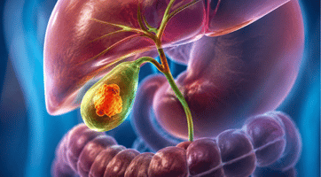
About Gallbladder Cancer Surgery
Expert Surgical Care for Gallbladder Cancer
Gallbladder cancer is an uncommon but aggressive cancer that often develops without noticeable symptoms in its early stages. Early diagnosis significantly improves the chances of successful treatment, with surgery offering the best opportunity for long-term disease control and cure.
Depending on the stage, location, and extent of the tumor, treatment may involve Robotic Radical Cholecystectomy or Open Radical Cholecystectomy. The procedure aims to achieve complete cancer removal with adequate lymph node clearance while preserving patient safety and ensuring the best possible oncological outcomes.
Radical Cholecystectomy (Robotic / Open)
Standard Surgical Treatment for Resectable Gallbladder Cancer
Radical Cholecystectomy is the standard surgical treatment for patients with resectable gallbladder cancer beyond the earliest stage. The procedure involves removal of the gallbladder along with a portion of the liver adjacent to the gallbladder and regional lymph nodes to ensure complete oncological clearance.
The operation may be performed using a robotic or open surgical approach depending on the stage of the tumor, anatomical complexity, and the individual patient's suitability.
Complete Removal of the Gallbladder
The cancerous gallbladder is completely removed to achieve optimal disease control.
Liver Wedge or Segment IVB & V Resection
Removal of adjacent liver tissue is performed whenever clinically indicated.
Regional Lymph Node Dissection
Nearby lymph nodes are removed for accurate staging and complete cancer clearance.
Removal of Involved Surrounding Tissues
Adjacent tissues affected by the tumor are removed whenever required for complete resection.
Reconstruction When Necessary
Reconstructive procedures are performed when required to restore normal digestive continuity.
Symptoms of Gallbladder Cancer
Recognize the Warning Signs
Gallbladder cancer is often difficult to detect in its early stages because symptoms may not appear until the disease has advanced. When symptoms do develop, they can resemble common conditions such as gallstones or a bile duct blockage. If these symptoms persist, prompt medical evaluation is recommended.
Upper Abdominal Pain
Persistent pain or discomfort in the upper right side of the abdomen.
Jaundice
Yellowing of the skin and the whites of the eyes due to bile duct blockage.
Abdominal Lump
A noticeable lump or swelling in the abdomen should be evaluated promptly.
Unexplained Weight Loss
Unexpected weight loss without changes in diet or physical activity.
Nausea & Vomiting
Persistent nausea or repeated vomiting may indicate gallbladder disease.
Bloating
Persistent abdominal bloating or a feeling of fullness after meals.
Fever
Recurring fever may occur, especially if infection or bile duct obstruction is present.
Stages of Gallbladder Cancer
Understanding How Gallbladder Cancer Progresses
Gallbladder cancer is classified into different stages based on how deeply the cancer has grown and whether it has spread to nearby lymph nodes, organs, or distant parts of the body. Identifying the stage helps determine the most appropriate treatment plan and provides insight into the expected prognosis.
Stage 0 (Carcinoma in Situ)
Abnormal, precancerous cells are confined to the inner (mucosal) lining of the gallbladder and have not invaded deeper tissues.
Stage 1
Cancer has developed in the mucosal layer and may extend into the muscle wall of the gallbladder.
Stage 2
The cancer has spread beyond the muscle layer into the connective tissue surrounding the gallbladder.
Stage 3
Cancer has spread to the liver, nearby organs, the outer (serosal) layer of the gallbladder, and may involve nearby lymph nodes.
Stage 4
Cancer has spread to multiple nearby lymph nodes, major blood vessels, or distant organs beyond the gallbladder.
Risk Factors for Gallbladder Cancer
Factors That Increase the Risk of Gallbladder Cancer
Several factors can increase the likelihood of developing gallbladder cancer. While having one or more of these risk factors does not necessarily mean a person will develop cancer, awareness can help with early detection and timely medical care.
Gallstones
Gallstones are the most significant risk factor for gallbladder cancer. Long-term irritation and chronic inflammation caused by gallstones may contribute to cancer development.
Age & Gender
Gallbladder cancer is more common in older adults, and women have a higher risk of developing the disease than men.
Ethnicity
The disease is seen more frequently in certain populations, including Native Americans, Hispanics, and people of South Asian descent.
Porcelain Gallbladder
A rare condition in which the gallbladder wall becomes calcified (covered with calcium deposits), increasing the risk of gallbladder cancer.
Chronic Gallbladder Inflammation
Long-standing inflammation of the gallbladder, such as chronic cholecystitis, may increase the risk of developing gallbladder cancer.
Important Note
Gallbladder cancer is a rare but serious disease. Early symptoms are often absent or mistaken for common gallbladder conditions, which means the disease is frequently diagnosed at a later stage. If you experience persistent abdominal pain, jaundice, unexplained weight loss, or other concerning symptoms, consult a healthcare provider promptly for evaluation and appropriate treatment.
Diagnosis & Tests for Gallbladder Cancer
How Gallbladder Cancer Is Diagnosed
Diagnosing gallbladder cancer involves a combination of blood tests, tumor marker tests, imaging studies, and surgical procedures. These tests help confirm the diagnosis, determine the extent of the disease, and guide the most appropriate treatment plan.
Liver Function Test (LFT)
Measures substances released by the liver to evaluate its function. Abnormal results may indicate that gallbladder cancer is affecting the liver or bile ducts.
CEA Blood Test
The Carcinoembryonic Antigen (CEA) assay measures a tumor marker in the blood. Elevated levels may suggest gallbladder cancer or other cancers.
CA 19-9 Assay
This blood test measures the CA 19-9 tumor marker. High levels may indicate gallbladder cancer, pancreatic cancer, or other biliary diseases.
Imaging Tests
Imaging studies help identify tumors and determine their location and spread. These include Abdominal Ultrasound, CT Scan, MRI, Endoscopic Ultrasound (EUS), and Endoscopic Retrograde Cholangiopancreatography (ERCP).
Surgical Diagnostic Procedures
In some cases, minimally invasive surgical procedures are required to obtain tissue samples or determine how far the cancer has spread.
Biopsy
A small tissue sample is removed and examined under a microscope. A biopsy is the only definitive method to confirm a diagnosis of gallbladder cancer.
Laparoscopy
A laparoscope (a thin, lighted tube) is inserted through a small incision to examine the gallbladder and nearby organs. This procedure helps determine whether the cancer has spread and assists in treatment planning.