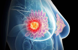
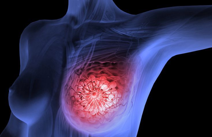
Breast cancer develops when cells within the breast begin dividing abnormally and form a tumour. Regular screening and prompt evaluation of any new symptom can improve the chances of detecting breast cancer early, when treatment tends to be more effective and less extensive.
The two most common forms are invasive ductal carcinoma, which starts in the milk ducts before spreading into surrounding breast tissue, and invasive lobular carcinoma, which begins in the lobules. Both can spread beyond the breast if left untreated, which is why treatment planning starts with confirming the exact type, stage and receptor status (ER, PR, HER2) through biopsy and imaging.
Risk Factors
Understanding Breast Cancer Risk Factors
Having one or more risk factors does not mean breast cancer is inevitable, and many women who develop it have no identifiable risk factor at all. Known contributors include increasing age (most cases are diagnosed after 50), inherited BRCA1 or BRCA2 mutations, a family history of breast or ovarian cancer, dense breast tissue, early onset of periods or late menopause, long-term hormone replacement therapy, limited physical activity, obesity after menopause, and regular alcohol consumption. Reproductive history — including a first pregnancy after 30, no full-term pregnancy, or not breastfeeding — may also play a role.
How Breast Cancer Is Diagnosed
Tests Used to Evaluate Breast Abnormalities
A breast ultrasound uses sound waves to examine areas of concern, while a diagnostic mammogram provides a more detailed X-ray when a lump or abnormal screening result requires further evaluation. Breast MRI may be used in selected cases to provide a more detailed view using magnetic imaging.
If any of these tests raise a concern, a biopsy — such as fine-needle aspiration, core biopsy or open biopsy — may be performed to confirm the diagnosis by examining tissue directly under a microscope. Being referred to a breast specialist for these tests does not, on its own, mean that cancer has been diagnosed.
Kinds of Breast Cancer
The Most Common Types of Breast Cancer
The most common kinds of breast cancer are Invasive Ductal Carcinoma (IDC) and Invasive Lobular Carcinoma (ILC). Both types are invasive cancers, meaning they can spread beyond the breast tissue to other parts of the body if left untreated.
Invasive Ductal Carcinoma (IDC)
The cancer cells begin in the ducts and then grow outside the ducts into other parts of the breast tissue. These invasive cancer cells can also spread, or metastasize, to other parts of the body.
Invasive Lobular Carcinoma (ILC)
Cancer cells begin in the lobules and then spread from the lobules to the nearby breast tissues. These invasive cancer cells can also spread to other parts of the body.
Conditions Treated
Comprehensive Breast Cancer and Breast Disease Care
A range of breast cancers, breast conditions and related treatment needs can be assessed individually to determine the most appropriate management and surgical approach.
Breast Cancer (All Stages)
Assessment and surgical management of breast cancer across different stages.
Ductal Carcinoma In Situ (DCIS)
Evaluation and treatment of non-invasive breast cancer confined to the milk ducts.
Invasive Breast Cancer
Surgical treatment of invasive breast cancers based on tumour type, stage and individual factors.
Recurrent Breast Cancer
Assessment and treatment planning for breast cancer that has returned after previous treatment.
Male Breast Cancer
Specialised assessment and surgical management of breast cancer in men.
Benign Breast Lumps
Evaluation and management of benign breast lumps, fibroadenoma and phyllodes tumours.
High-Risk Breast Lesions
Assessment and appropriate management of breast lesions associated with an increased cancer risk.
Chemoport Placement and Removal
Placement and removal of chemoports to provide reliable venous access during cancer treatment.
Breast Cancer Treatment Options
Individualised Treatment Based on Cancer Type and Stage
The right combination of treatments depends on tumour size, stage, hormone receptor status and the patient's overall health. Treatment is planned individually to provide appropriate cancer control while considering functional and cosmetic outcomes.
Lumpectomy (Breast-Conserving Surgery)
Removes the tumour along with a margin of healthy tissue, usually for smaller tumours. Some larger tumours can be shrunk with chemotherapy beforehand to make breast-conserving surgery possible.
Mastectomy
Removes the entire breast. Skin-sparing and nipple-sparing techniques may improve cosmetic outcomes for selected patients.
Sentinel Lymph Node Biopsy and Axillary Dissection
Sentinel node biopsy checks the first lymph nodes that the tumour drains into. If cancer is found there, further lymph node removal from the armpit may be recommended.
Radiation, Hormone, Targeted and Immunotherapy
Radiation therapy targets residual cancer cells with high-energy beams. Hormone therapy is used for ER/PR-positive cancers, targeted therapy for HER2-positive cancers, and immunotherapy may be an option in selected triple-negative cases. The appropriate treatment depends on the tumour's individual biology.
Diagnosis of Breast Cancer
Tests Used to Detect and Confirm Breast Cancer
Doctors often use additional tests to find or diagnose breast cancer. They may refer women to a breast specialist or a surgeon. This does not mean that she has cancer or that she needs surgery. These doctors are experts in diagnosing breast problems.
Breast Ultrasound
A machine that uses sound waves to create pictures, called sonograms, of areas inside the breast.
Diagnostic Mammogram
If you have a breast problem, such as a lump, or if an abnormal area is seen on a screening mammogram, your doctor may recommend a diagnostic mammogram, which is a more detailed X-ray of the breast.
Breast MRI
Breast Magnetic Resonance Imaging (MRI) uses a powerful magnet connected to a computer to create detailed images of areas inside the breast.
Biopsy
A biopsy removes tissue or fluid from the breast for examination under a microscope. Different types include fine-needle aspiration, core biopsy, and open biopsy.
Conditions Treated
Comprehensive Breast Cancer & Breast Surgery Services
Dr. Tapan Singh Chauhan provides expert surgical management for breast cancer, benign breast diseases, and chemoport procedures using evidence-based surgical techniques. Every treatment plan is personalized according to the patient's diagnosis, stage of disease, and overall health to achieve the best possible outcomes.
Breast Cancer
Benign Breast Lumps
Fibroadenoma
Phyllodes Tumor
Ductal Carcinoma In Situ (DCIS)
Invasive Breast Cancer
Recurrent Breast Cancer
Male Breast Cancer
High-Risk Breast Lesions
Chemoport Placement
Chemoport Removal
Long-Term Venous Access for Chemotherapy
Chemoport
Safe & Comfortable Venous Access for Chemotherapy
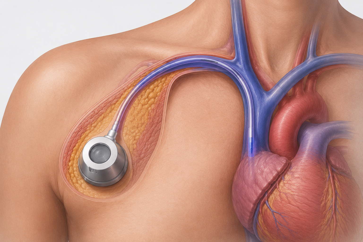
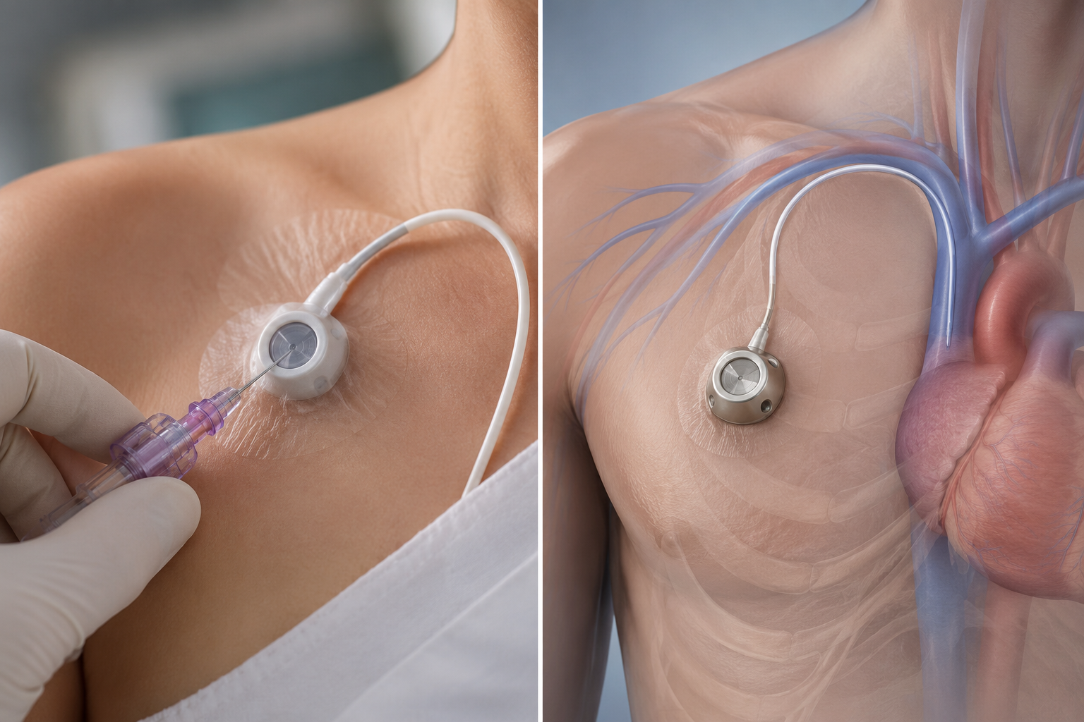
A Chemoport (also called an implantable venous access port) is a small medical device placed beneath the skin and connected to a large vein through a thin catheter. It provides secure, reliable, and long-term venous access for chemotherapy, targeted therapy, immunotherapy, blood sampling, blood transfusions, and other intravenous medications without repeated needle pricks in the arm.
The procedure is performed under local or general anesthesia through a small incision. The port remains completely beneath the skin, allowing patients to carry out their daily activities comfortably. It can safely remain in place for months or even years until cancer treatment is completed and can be removed once it is no longer required.
Why is a Chemoport Recommended?
Safe, Comfortable & Reliable Venous Access for Chemotherapy
A chemoport is a small implantable device placed beneath the skin that provides long-term venous access for patients undergoing chemotherapy. It makes treatment safer, more comfortable, and more convenient by reducing repeated needle punctures and protecting the veins throughout the course of cancer treatment.
Avoids Repeated Needle Pricks
Provides a single long-term access point, eliminating the need for frequent needle insertions.
Safe Delivery of Chemotherapy
Allows chemotherapy drugs to be administered safely into a large central vein.
Reduces Vein Damage
Protects small peripheral veins from irritation and damage caused by chemotherapy drugs.
Comfortable Long-Term Venous Access
Provides reliable access for prolonged treatment with greater patient comfort.
Allows Blood Sampling Through the Same Port
Blood tests can often be performed through the chemoport, reducing additional needle sticks.
Lower Risk of Vein Irritation
Helps prevent inflammation, pain, and irritation associated with repeated intravenous chemotherapy.
Improves Patient Convenience During Treatment
Makes repeated chemotherapy sessions easier and more comfortable while supporting a better treatment experience.
Recovery After Surgery
Personalized Postoperative Care for Faster Recovery
Recovery after breast cancer surgery or chemoport procedures depends on the type of surgery performed and the patient's overall health. Dr. Tapan Singh Chauhan provides comprehensive postoperative care, helping patients recover safely, minimize complications, and return to their daily activities as early as possible.
Early Mobilization
Effective Pain Management
Detailed Wound Care Guidance
Regular Follow-up Visits
Rehabilitation When Required
Long-Term Surveillance for Cancer Recurrence
Frequently Asked Questions
Find answers to common questions about breast cancer surgery, chemoport placement, treatment options, recovery, and surgical care.